Julie Hamilton Elliott, Cardiology Specialist at Northern Ireland Veterinary Specialists, recently implanted an artificial pacemaker for Ceaser, a 12-year-old Doberman Pinscher.
Julie is currently the only EBVS Specialist Cardiologist offering interventional cardiac procedures in Ireland, making NiVS the only specialist-led centre offering this lifesaving procedure to pets within the country.
Ceaser presented to his primary vet for severe lethargy and exercise intolerance. Investigations revealed high grade 2nd degree AVB with an average heart rate of 30bpm.
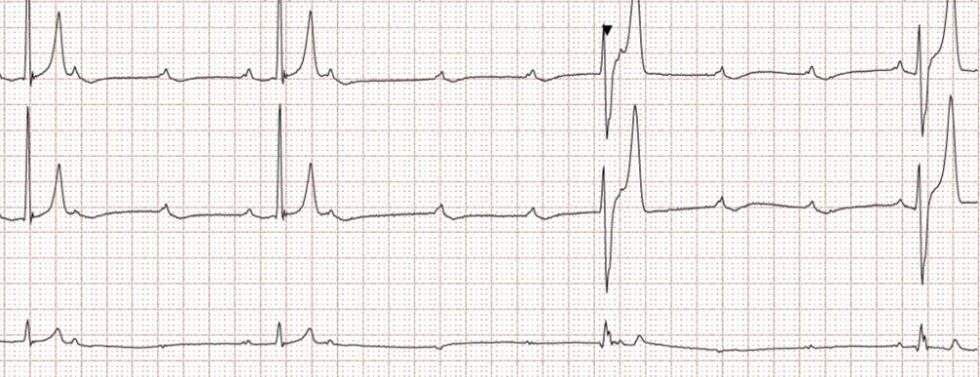
Holter recording showing sinus beats with multiple unconducted P waves and ventricular escape beats, consistent with high grade 2nd degree AVB.
Ceaser was referred to NiVS and underwent various tests which ruled out secondary causes, such as structural heart disease and systemic disease such as neoplasia. With no underlying treatable cause, medical management of high grade 2nd degree AVB is often unrewarding. Risks include progression to 3rd degree AVB and sudden cardiac death. Therefore, a permanent artificial pacemaker was recommended.
Artificial pacemaker implantation in dogs is usually performed transvenously as a minimally invasive procedure under general anaesthetic. The pacemaker lead is inserted in the right jugular vein and fluoroscopically guided to the apex of the right ventricle. After attaching a pulse battery to the lead and testing pacemaker function, a small subcutaneous pocket is created for the battery. Throughout the procedure, cardiac rhythm and output is monitored and external pacing patches often applied as a ‘back up’ if the heart rate becomes dangerously low.
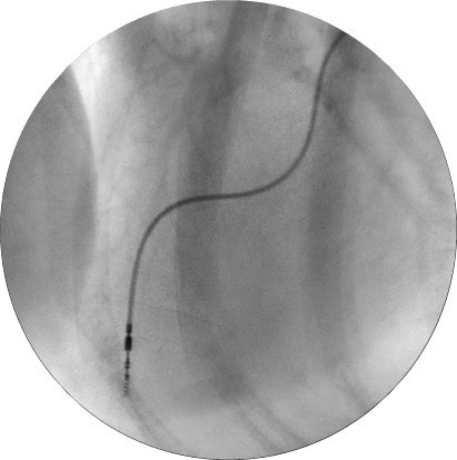
Fluoroscopic image of the heart showing the pacemaker lead in the right ventricle.
Ceaser rested for 4 weeks to reduce the risk of lead dislodgement. He experienced no complications and after his 1-month post-operative check the pacemaker settings were adjusted to allow his heart rate to increase with movement. He can now exercise and is back to his normal self.
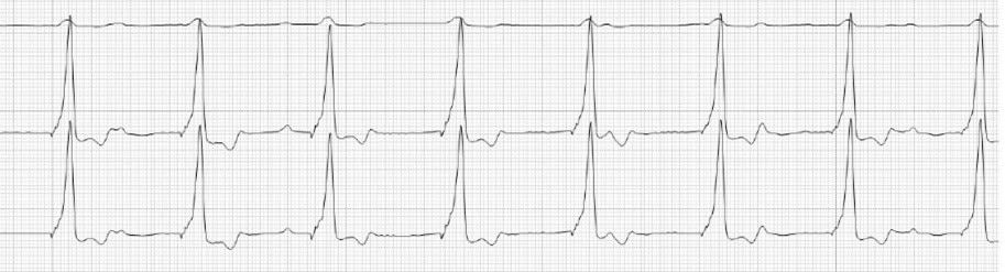
Post-operative ECG showing pacemaker rhythm. Beats appear as regular ventricular beats due to the positioning within the right ventricle. Occasional sinus P waves can be seen.