Julie Kavanagh, Specialist in Veterinary Cardiology, shares tips on how to determine the significance of puppy murmurs and why early diagnosis can be life-changing.
A heart murmur indicates turbulent blood flow through the heart or its surrounding vessels. Whilst echocardiography is required for definitive diagnosis, the characteristics of a murmur (intensity, location and timing), together with the breed, may allow for a high index of suspicion as to the underlying cause.
Describing a heart murmur
There are 3 parts to the standard description of a heart murmur.
- Intensity is described as grade 1-6/6 based on the following descriptions;
Grade I: Very soft, focal murmur, heard only after a period of listening.
Grade 2: Readily appreciated but focal. Intensity less than heart sounds.
Grade 3: Intensity equal to heart sounds.
Grade 4: Intensity greater than heart sounds but no palpable precordial thrill.
Grade 5: Palpable precordial thrill.
Grade 6: Palpable precordial thrill and murmur still audible when stethoscope lifted 1-2mm off chest. - Location describes the region at which you hear the murmur loudest (point of maximal intensity). A heart murmur can be localised to the left or right side of the thorax, and the cardiac base (towards the head) or apex (towards the tail.)
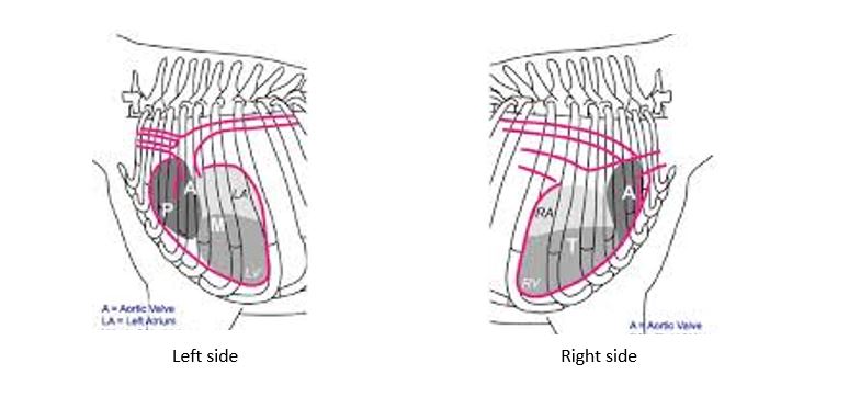
Consider the cardiac valves and vessels that occur in these regions;
Left base: Aorta and aortic valves, Pulmonary artery and pulmonic valves
Left apex: Mitral valve
Right Base: Ascending aorta
Right Apex: Tricuspid valve
Consider the common congenital defects that can affect these structures (below)
Timing of a heart murmur describes the part of the cardiac cycle in which it is occurring. Most heart murmurs occur during systole (between the first and second heart sounds) and are described as systolic. Mitral and tricuspid valve regurgitation causes a systolic murmur. Continuous murmurs occur throughout the entire cardiac cycle (both systole and diastole) with no break. Diastolic murmurs occur after the second heart sound and are very rare. Aortic and pulmonic valve insufficiency causes diastolic murmurs.
Left-sided heart murmurs
Patent ductus arteriosus (PDA)
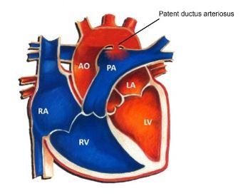
PDA describes a communication between the aorta and pulmonary artery. Because the blood pressure within the aorta is higher that the pulmonary artery in both systole and diastole, blood continuously shunts from the aorta to pulmonary artery and this causes a continuous heart murmur. The location of a PDA is the left heart base. Sometimes the murmur is only apparent very cranially, under the axilla so be sure to move the left forelimb forward when auscultating a puppy.
Systemic diastolic blood pressure is reduced, because some blood escapes from the aorta in to the pulmonary artery. This causes a wider pulse pressure (the difference between systolic and diastolic pressure) which is appreciated on the clinical exam as hyperdynamic or bounding pulses.
Summary
PDA heart murmur features:
- Any grade (but usually >grade 4)
- Location at left heart base (or more cranial)
- Continuous
Additional clinical exam findings:
- Hyperdynamic (bounding) pulses
PDA Breeds:
- Maltease
- Collie
- German Shepherd
- Chihuahua
- Corgi
PDA is the only congenital heart disease that is completely curable if identified and closed early. Failure to do so can result in congestive heart failure or over-circulation of the lungs leading to pulmonary hypertension with reversal of PDA flow and cyanosis. Left untreated, most dogs with a PDA will die before 1 year of age. A continuous heart murmur is never innocent and immediate investigation with echocardiography is indicated.
Pulmonic stenosis (PS)
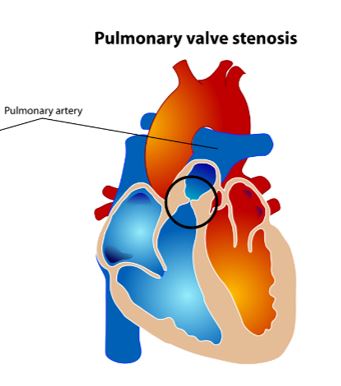
PS describes a ‘narrowing’ in the pulmonary artery (PA), usually at the level of the pulmonic valves. This causes obstruction to blow flood between the right ventricle and the lungs. The degree of obstruction is classified by echocardiography as mild, moderate or severe. Dogs with severe PS are at risk of right-sided congestive heart failure (jugular distention, pleural effusion and ascites), exercise intolerance or syncope.
Pulmonic balloon valvuloplasty is available for suitable patients with severe PS. This can reduce the obstruction across the PA and significantly alter prognosis.
PS heart murmur features:
- Any grade
- Location at left heart base
- Systolic
PS breeds:
- Bulldogs (French and English)
- Cocker Spaniels
Sub-aortic stenosis (SAS)
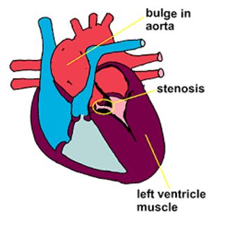
SAS describes a narrowing in the aorta, below the aortic valves. This causes obstruction to blow flood between the left ventricle and the body.
Like with PS, the degree of obstruction is classified by echocardiography as mild, moderate or severe. Dogs with severe SAS are at risk of arrhythmias and sudden death, less commonly they may develop left-sided congestive heart failure (pulmonary oedema).
SAS is present at birth but, unlike PS, SAS can worsen over the first year of life so serial echos may be required for accurate diagnosis. Medication (beta-blockers) may be prescribed to moderate and severe cases. Aortic balloon valvuloplasty may be considered in very severe cases.
Because of the path of the aorta, a murmur associated with SAS may also be appreciated at the left thoracic inlet.

SAS heart murmur features:
- Any grade
- Location at left heart base (may be appreciated at thoracic inlet also)
- Systolic
SAS breeds:
- Golden Retrievers
- Boxers
Mitral valve dysplasia (MVD)
MVD occurs when the mitral valves (MV) develop abnormally. This can result in the valve being too narrow (stenotic) and/or incompetent (regurgitation).
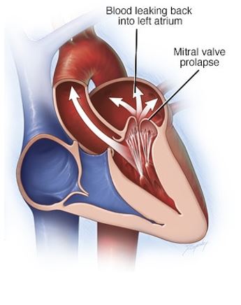
The MV should be open during diastole, allowing the left ventricle to fill with blood. If it is stenotic, this will cause turbulence to blood flow entering the LV and result in a diastolic murmur.
The MV should be closed during systole, allowing blood to be pumped from the LV to the body via the aorta. If the MV is incompetent, some blood will regurgitate back into the left atrium. This will cause a systolic murmur.
MVD heart murmur features:
- Any grade
- Location at Left Apex
- Systolic and/or diastolic. Presence of both causes a ‘to-and-fro’ murmur.
MVD breeds:
- Labrador
- English Bull Terriers
- English Springer Spaniels
Right-sided heart murmurs:
Ventricular septal defect (VSD)
 VSD describes an abnormal communication between the left and right ventricles, representing abnormal foetal development. Typically this results in shunting of blood from the high pressure LV into the lower pressure RV during systole. The significance of a VSD can only be determined by echocardiography and is largely based on the size of the defect. Small defects are described as ‘restrictive’. They are not haemodynamically significant and no treatment is required. When the defect is restrictive, blood shunts through it very quickly resulting in turbulence in the RV and a loud heart murmur. This is a rare scenario in which a loud murmur may reflect mild disease.
VSD heart murmur features:
- Any grade (louder may be better!)
- Location on right side (base or apex)
- Systolic
VSD breeds:
- Border terriers
- Keeshound
- Cats!
Tricuspid valve dysplasia (TVD)
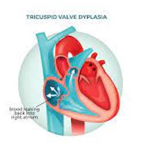
TVD occurs when the tricuspid valves (TV) develop abnormally. This can result in the valve being too narrow (stenotic) and/or incompetent (regurgitation).
The TV should be open during diastole, allowing the right ventricle to fill with blood. If it is stenotic, this will cause turbulence to blood flow entering the RV and result in a diastolic murmur. The TV should be closed during systole, allowing blood to be pumped from the RV to the lungs via the pulmonary artery. If the TV is incompetent, some blood will regurgitate back into the right atrium. This will cause a systolic murmur.
TVD heart murmur:
- Any grade
- Location at Right Apex
- Systolic
TVD breeds:
- Labrador
Innocent heart murmurs
‘Innocent’ or physiological heart murmurs occur in the absence of structural cardiac disease. A change in blood viscocity (eg anaemia or pyrexia) lowers the ‘threshold’ for turbulence formation and can result in an audible murmur.
Only echocardiography can exclude structural heart disease and therefore confirm a murmur is physiological.
Innocent heart murmur features:
- Always < grade 3
- Location variable but usually Left Base
- Always Systolic


