Cara – a six-year-old female neutered Cavalier King Charles Spaniel was referred to Northern Ireland Veterinary Specialist’s (NIVS) soft tissue surgery service for further assessment and treatment of a large hepatic mass. She had presented to her referring vets for a routine exam.
The referring vet very astutely noticed a firm and distended abdomen which would prompt further investigation. Routine bloods were performed which revealed a mild regenerative anaemia, elevated ALP and very elevated ALT. There was a high index of suspicion for a liver mass. Abdominal radiographs showed a large cranial abdominal mass and deviation of the intestines. Ultrasound confirmed the presence of a very large liver mass.
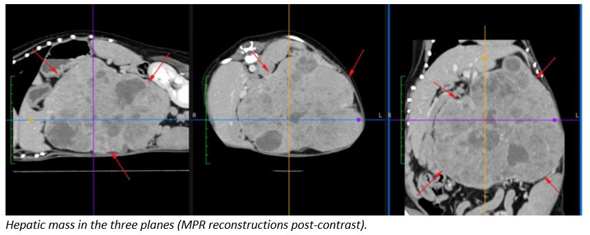
To better appreciate the 3-D anatomy and relationship with the surrounding structures, a CT was performed. The CT report confirmed a large heterogenous mass arising from the left side of the liver. The left lateral, left medial and papillary process of the caudate lobe were involved and some of the local lymph nodes enlarged. The mass was very suspicious for neoplasia.
On referral to our surgery service a clinical examination was performed by Surgery Specialist Aidan McAlinden. On physical exam she was quiet but alert and responsive. Her heart rate and respiratory rate were within normal limits. Her gums were slightly pale and periodontal disease present, mostly affecting the canines and premolars. No jaundice or dehydration was evident on exam. Peripheral lymph nodes were evaluated and noted to be of normal size. Severe abdomenomegaly was very apparent when walking in to the examination room. Her cranial abdomen was firm, uncomfortable and a mass was palpable on both sides of the abdomen just caudal to the costal arch. She was cachexic in appearance, with poor coverage of the dorsal spinous processes along the length of her spine.
The CT report and history was studied in detail by our Surgical Specialist Aidan. His most crucial finding was the presence of a left-sided hepatic mass. This has shown to be of positive prognostic significance especially in the case of Hepatocellular Carcinoma (HCC). Multiple studies have highlighted excellent outcomes in dogs with these left sided HCCs. This is most likely due to the left medial and lateral liver lobes being away from vital structures such as the portal vein, caudal vena cava and gallbladder. A massive morphology, as in this case, has shown to be favourable when compared to the diffuse and nodular forms. This finding ultimately comes down to resectability as nodular and diffuse forms may require removal of the entire liver. Massive forms are mostly localised. Other differentials included a Biliary Duct Carcinoma (uncommon), Carcinoid or primary sarcoma (very rare).
A very in-depth conversation was had with the owner highlighting possible outcomes and risks. It was advised that the tumour may be resectable although involving multiple lobes. Overall, prognosis would depend on histopathology, margin achieved and whether or not it has metastasized regionally. The owner was informed of significant perioperative risk of severe complications and potentially death. Owner was also made aware that if the mass was not resectable we may have to euthanise on the table. After due consideration it was decided to proceed with an exploratory celiotomy to determine resectability and hopefully achieve a positive outcome.
Due to the perioperative risk of haemorrhage her blood was typed. She was DEA 1.1 positive and canine packed red blood cells sourced. Clotting times were assessed and within normal limits. Haematology and biochemistry were repeated. There was a mild leucocytosis and mild non-regenerative anaemia. The PCV was 30% and platelets an adequate number. There was mild increase in bile acids and massive elevation of both ALT and ALP.
A ventral midline celiotomy was performed. A massive, multlobular left-sided hepatic mass occupying the whole cranial half of the abdomen was identified. This created a space occupying effect and displaced the right side of the liver, stomach, spleen and left kidney into the periphery. The mass was pressing against the right side of the liver, but it was not invading the papillary process of the caudate lobe.
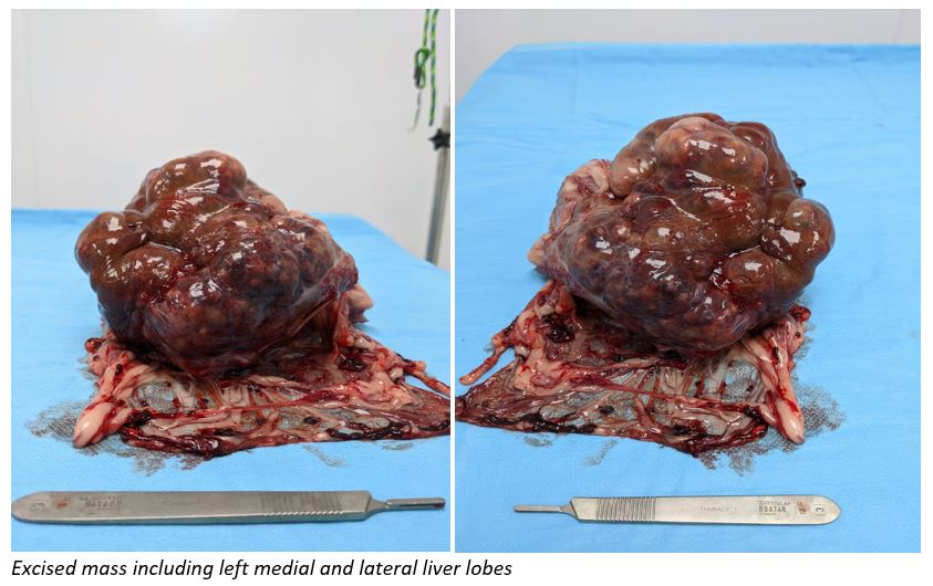
The mass was exteriorised and the hilus of the left medial and lateral lobes was isolated. A TA30-V3 stapler was applied across it. The liver was transected and was further elevated out of the abdomen. It was enshrouded in omentum and this was released circumferentially with the ligasure including en-bloc resection of the two enlarged hepatic nodes. Minor haemorrhage from the omentum was controlled with bipolar cautery. A 3-0 PDS ligature was placed around the hilus of the left side of the liver to encircle portal and hepatic branches. Surgicel was applied to the transected edge of the liver. The abdomen was lavaged and gloves and instruments were changed before closing in routine fashion. Multiple samples from the liver and lymph nodes were taken and submitted for histopathology.
She had a relatively unremarkable recovery and was eating food the following evening thanks to the fantastic nursing staff. Two days post-op she was discharged with histopathology results pending. She was delighted to be reunited with her Mommy after her big procedure.
Histopathology results were received a week later. The results confirmed a hepatocellular carcinoma as predicted and there was no evidence of metastasis to the regional lymph nodes. Overall a favourable prognosis was provided. Although at first glance this case may have appeared hopeless, with a quick review of the literature and a competent, steady surgical hand, an excellent outcome can be achieved. The owner advised us that she is doing very well and is almost back to her normal self. All of us here at NIVS were delighted to receive the positive news and that she has recovered so well. We wish them plenty of happy memories for years to come!